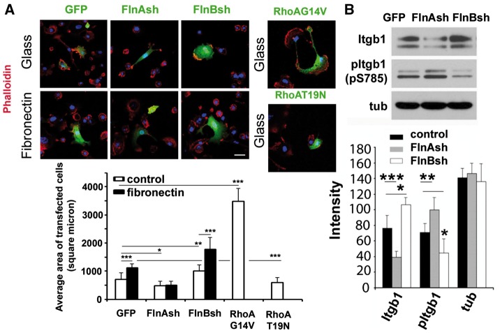
Figure 6.
β1 integrin dependent activation of RhoA via FlnA/B regulates cell spreading. (A) Fluorescence photomicrographs demonstrate that fibronectin stimulation causes increased cell spreading in wildtype primary mouse chondrocytes. Cell spreading is diminished in FlnA knockdown primary mouse chondrocytes, regardless of fibronectin stimulation, consistent with impairment in RhoA activation. Conversely, increased cell spreading is seen with FlnB inhibited cells, following plating on either glass or fibronectin. FlnB knockdown cells display increased activated RhoA expression levels (see Fig. 5). Transient transfection of activated RhoA (G14V) leads to a similar pattern of spreading as seen with FlnB inhibition. Similar to the FlnA null cells and compared to constitutively active RhoA transfected cells, transfection of inactive RhoA (T19N) leads to impaired cell spreading. Changes in cell spreading area are quantified graphically for the different experimental variables. (B) Western blot demonstrates that loss of FlnA impairs β1-integrin expression levels but promotes expression of phospho- β1-integrin at serine 785. Phospho-integrin β1 (S785) promotes cell attachment but prevents spreading and migration (26). The opposite changes are seen with FlnB knockdown. *P≤0.05, **P≤ 0.01, ***P ≤ 0.001. Scale bar = 25 μm.

