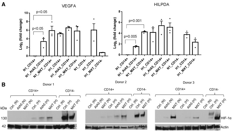
Figure 4.
AtA5 in normoxia induces hypoxic gene expression in monocytes without robust stabilization of HIF-1α. (A) Bar graph depicts fold changes in VEGF and HILPDA gene expression in normoxic and hypoxic CD14+ and CD14– cells upon treatment with AtA5 or MXT for 24 h (1 day) (n = 3 donors). (B) Immunoblot shows the expression of HIF-1α in lysates (40 µl) of CD14+ and CD14– cells examined in (A). The cells were isolated from PBMCs at room conditions followed by culture (5–7 million/ml) for 24 h in normoxia or hypoxia (1%) upon treatment with AtA5 or MXT. Actin was used as a loading control.

