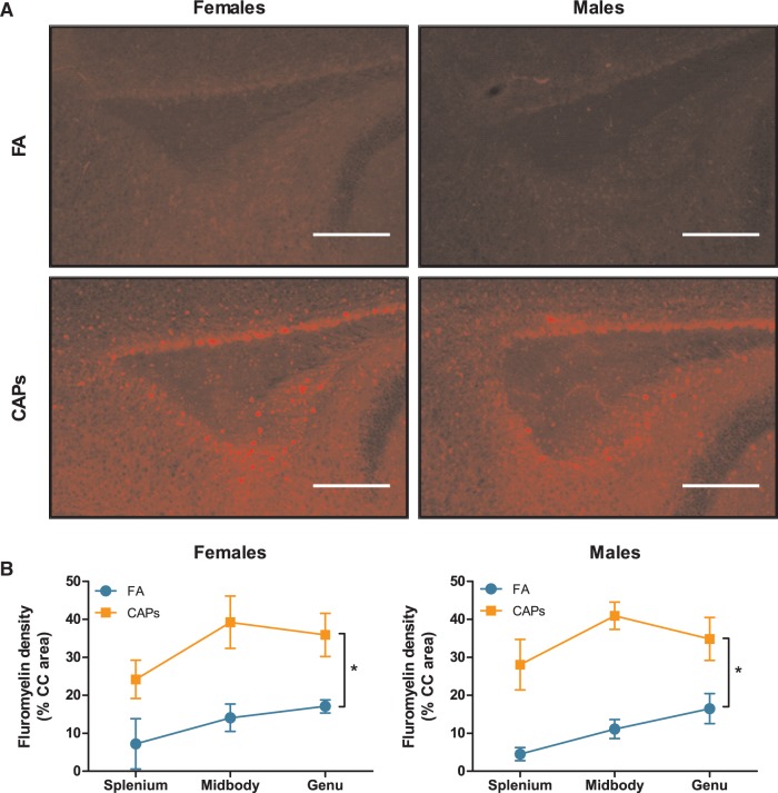
FIG. 6.
Prenatal CAPs exposure results in increased presence of compact myelin in the CC. CAPs significantly increased compact myelin, as indicated by FluoroMyelin Red staining density, across the 3 major regions indicated: genu (rostral portion), midbody (central portion), and splenium (caudal portion). (A) Representative microcraphs of PND15 splenium at 10X magnification. Scale bars represent 500μm distance. B) Data is expressed as a percent of CC area positively stained by FluoroMyelin. Statistical outcome: * = main effect of CAPs across sexes; main effect of PND. Data represent mean ± SEM of 3 serial sections of brain tissue (N = 6–11 per group).

