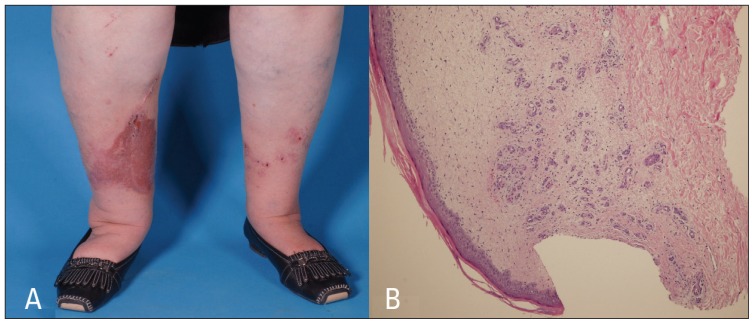
Figure 1.
A) Multiple well-defined erythematous to violaceous indurated scaly papules and plaques with some excoriated papules on both lower legs (more in right leg, at the site of the surgical scar). B) Lobular proliferation of small capillaries in a loose stroma with extravasated erythrocytes and a sparse mononuclear cell infiltrate throughout the upper dermis. Interstitial fibroblast cells are slightly increased in number. Hyperkeratosis and mild spongiosis of the overlying epidermis. (Hematoxylin eosin stain 1×10).

