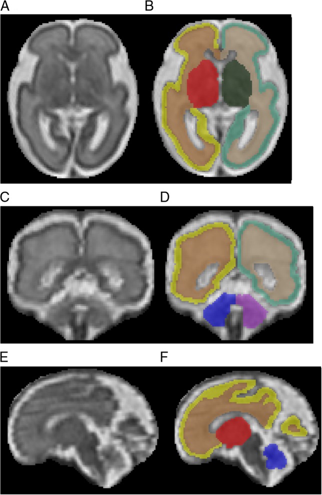
Figure 1.

T2-Weighted of the high-resolution fetal brain reconstruction at 27 weeks gestation in the axial (A and B), coronal (C and D), and sagittal (E and F) planes, with anatomical images on the left (A, C, E) and corresponding segmentations on the right (B, D, F). Color key: yellow = left CGM; brown = left FWM; red = left DSS; blue = left cerebellum; light green = right CGM; tan = right FWM; dark green = right DSS; purple = right cerebellum.
