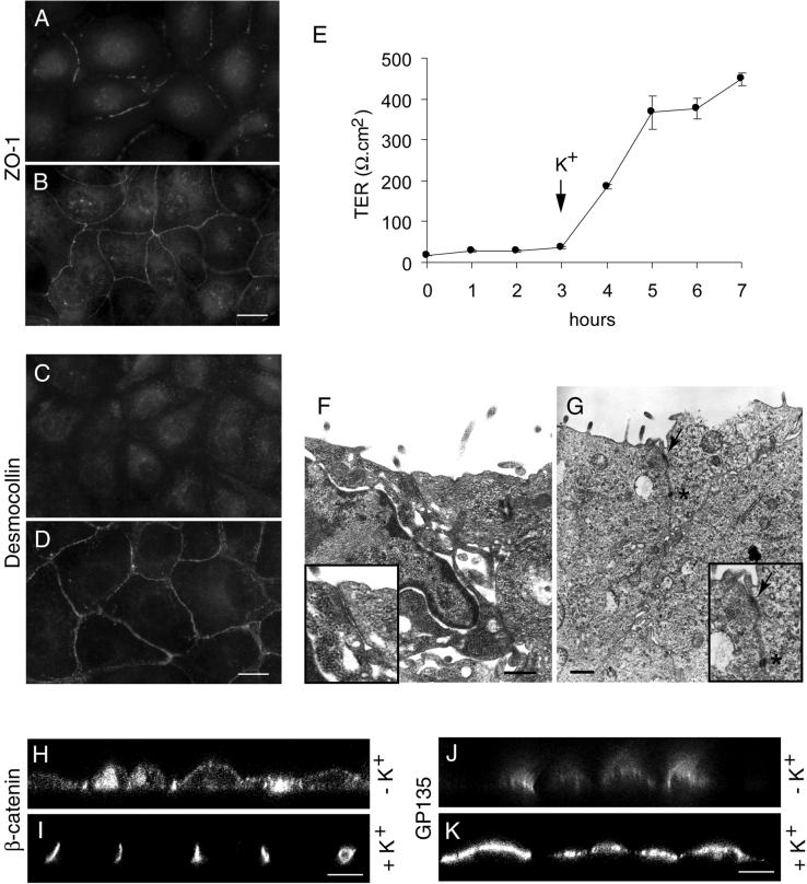
Figure 2.
Na,K-ATPase-mediated inhibition of formation of tight junctions and desmosomes is reversible. Immunofluorescence localization of ZO-1 (A and B) and desmocollin (C and D). Cells incubated in K+-free medium show an incomplete ZO-1 ring at the plasma membrane region (A) and intracellular localization of desmocollin (C). Replenishment of K+ results in a complete ZO-1 ring (B) and plasma membrane localization of desmocollin (D). (E) Measurement of TER. Note an increase in the TER as soon as the cells are shifted to K+-containing medium. (F and G) Transmission electron microscopy. Note the absence of tight junctions (arrow) and desmosomes (asterisk) in K+-free medium (F) and their presence in K+-containing medium (G). Insets in F and G are the higher magnifications of tight junction regions. (H–K) Confocal microscope X-Z sections. Note the nonpolarized distribution of β-catenin (H) and GP135 (J) under K+-depleted condition and the polarized distribution of β-catenin (I) and GP135 (K) after K+ repletion. Bars in A–D, 10 μm; F and G, 500 nm; and H–K, 5 μm.

