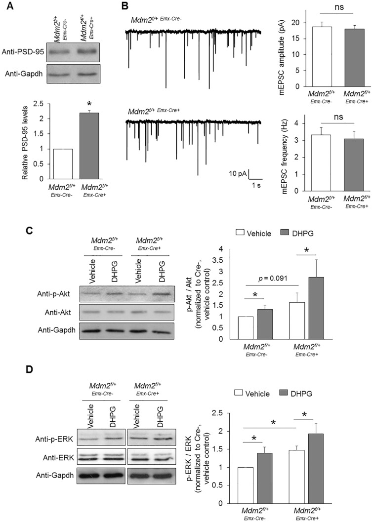
Figure 5.
Knocking down Mdm2 does not alter synaptic function or Gp1 mGluR signaling. (A) Representative western blots of PSD-95 and Gapdh, and quantification from Mdm2f/+-Emx-Cre+ or Mdm2f/+-Emx-Cre-cortical neuron cultures (n = 8). (B) Patch-clamp recording from Mdm2f/+-Emx-Cre+ or Mdm2f/+-Emx-Cre-cortical neurons at DIV 14. Representative mEPSC traces, and quantification of mEPSC amplitude and frequency are shown (n = 21 for both Mdm2f/+-Emx-Cre+ and Mdm2f/+-Emx-Cre-neurons). (C, D) Representative western blots of Akt, p-Akt, ERK, p-ERK and Gapdh, and quantification from Mdm2f/+-Emx-Cre+ or Mdm2f/+-Emx-Cre-cortical neuron cultures after vehicle or DHPG treatment for 30 min (n = 7 and 6 for (C) and (D), respectively). For the quantification above, a one-sample t-test (A), Student t-test (B) or a two-way ANOVA with Tukey test (C, D) was used. Data are represented as mean ± SEM with *P < 0.05, **P < 0.01, ns: non-significant.

