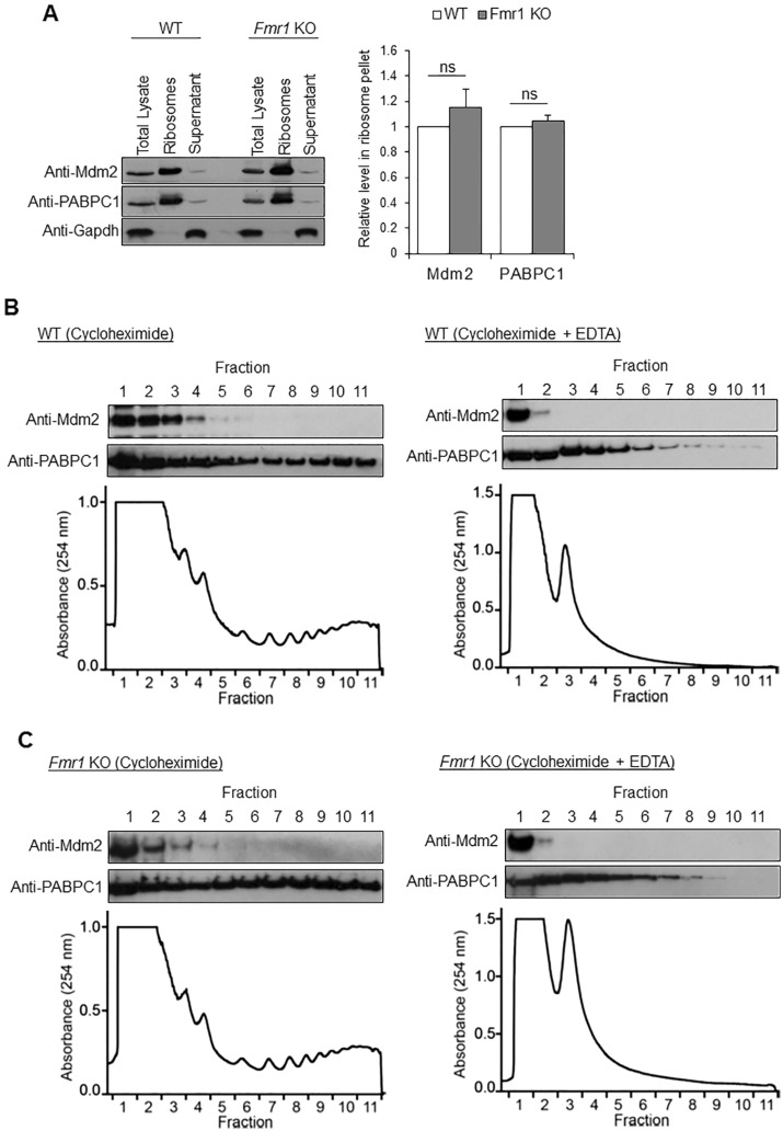
Figure 6.
Mdm2 associates with ribosomes. (A) Representative western blots of Mdm2, poly(A) binding protein cytoplasmic 1 (PABPC1) and Gapdh after sucrose density ultracentrifugation of WT or Fmr1 KO whole brains. PABPC1 and Gapdh serve as positive and negative controls, respectively, for ribosomes. Quantification is shown on the right (n = 4). (B, C) Representative western blots of Mdm2 and PABPC1, and polysome analysis from WT (B) or Fmr1 KO (C) brains. EDTA was used as a control to validate the fractionation by disrupting ribosomes.

