Figure 3.
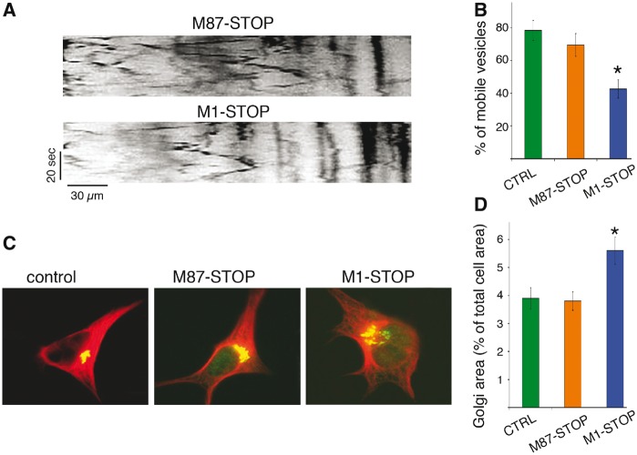
Abnormal transport and distribution of organelles in SH-SY5Y cells expressing M1-STOP spastin. SH-SY5Y cells stably expressing M1-STOP and M87-STOP spastins (see Supplementary Materials) were transfected with plasmids encoding an RFP-tagged version of synaptophysin (RFP-syn). The proportion of RFP-syn-positive mobile vesicles in neurites was analyzed using live-cell imaging, and representative kymographs are shown in (A) for comparison. (B) Compared to control (ctrl, GFP-transfected) cells, M87-STOP cells showed similar fraction of mobile RFP-syn-positive vesicles (78.3±6.2% vs 69.4 ± 5.6%). In contrast, M1-STOP cells showed a marked reduction in the proportion of these vesicles (42.5±5.5%). (C) Representative images of SH-SY5Y cells showing immunoreactivity for the cis Golgi marker GM130 (in green) and tubulin (in red). (D) Quantitation of GM1-130 immuno-positive area/total cell area revealed a wider distribution of the Golgi apparatus in M1-STOP cells (5.6±0.38%), compared to both control (3.9±0.38%) and M87-STOP cells (3.8±0.49% respectively). *P < 0.05. Bar, 10 microns.

