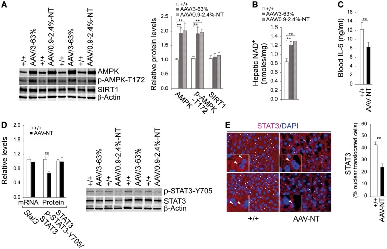
Figure 1.
Analysis of hepatic AMPK, SIRT1 and STAT3 signaling in 66–88 week-old wild-type and rAAV-treated G6pc-/- mice. For quantitative RT-PCR, the data were analyzed for wild-type (+/+, n = 13) and AAV-NT (n = 22) mice. (A) Western-blot analysis of AMPK, p-AMPK-T172, SIRT1 and β-actin with quantification of protein levels by densitometry in wild-type (n = 8), AAV/3–63% (n = 4) and AAV/0.9–2.4%-NT (n = 4) mice. (B) Hepatic NAD+ levels in wild-type (n = 13), AAV/3–63% (N = 8), and AAV/0.9–2.4%-NT (n = 8) mice. (C) Blood IL-6 levels in wild-type (n = 13) and AAV-NT (n = 16) mice. (D) Quantification of Stat3 mRNA by real-time RT-PCR; Western blot analysis of p-STAT3-Y705, STAT3 and β-actin with quantification of protein levels by densitometry from 4 pairs of wild-type/AAV-NT mice. The AAV-NT analyzed included AAV/3–63% (n = 2) and AAV/0.9–2.4%-NT (n = 2) mice. (E) Immunofluorescence analysis of hepatic STAT3 nuclear localization and quantification of nuclear STAT3-translocated cells. Representative plates shown are analyzed for wild-type mice (n = 6) and AAV-NT (n = 5) mice. Scale bar: 25 μm. Arrows denote STAT3-positive nuclei. Data represent the mean ± SEM; **P < 0.005.

