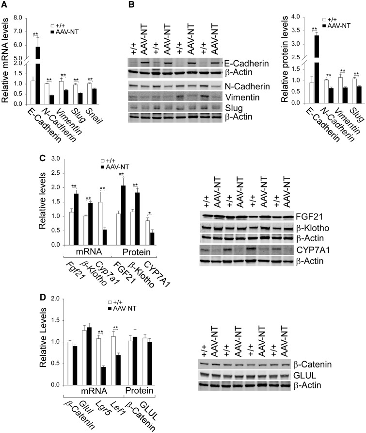
Figure 3.
Analysis of hepatic E-cadherin, mesenchymal markers, FGF21, β-klotho and β-catenin targets in 66–88 week-old wild-type and AAV-NT mice. For quantitative RT-PCR, the data were analyzed for wild-type (+/+, n = 13) and AAV-NT (n = 22) mice. For densitometry analysis, the data were analyzed from 4 pairs of wild-type/AAV-NT mice. The AAV-NT analyzed included AAV/3-63% (n = 2) and AAV/0.9–2.4%-NT (n = 2) mice. (A) Quantification of E-cadherin, N-cadherin, vimentin, Slug and Snail mRNA by real-time RT-PCR. (B) Western blot analysis of E-cadherin, N-cadherin, vimentin, Slug and β-actin with quantification of protein levels by densitometry. (C) Quantification β-Klotho, Fgf21 and Cyp7a1 mRNA by real-time RT-PCR; Western blot analysis of FGF21, β-klotho, CYP7A1 and β-actin; and quantification of protein levels by densitometry. (D) Quantification of β-Catenin, Glul, Lgr5, Cyclin D1 and Lef1 mRNA by real-time RT-PCR; Western-blot analysis of β-catenin, GLUL and β-actin; and quantification protein levels by densitometry. Data represent the mean ± SEM; *P < 0.05; **P < 0.005.

