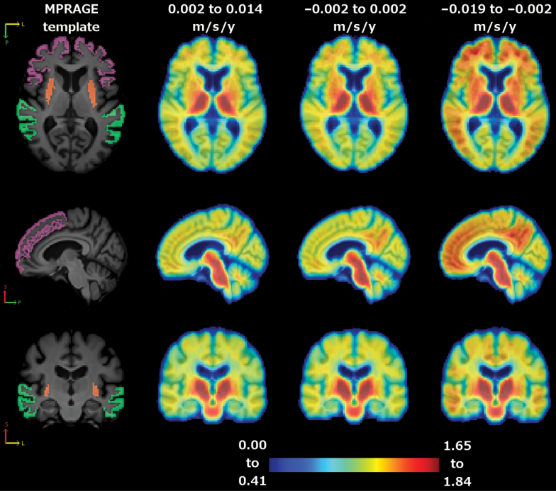
Figure 2.
Average cortical distribution volume ratio (cDVR) images by tertile of gait decline. Left to right: magnetization-prepared rapid gradient echo (MPRAGE) template (orange: putamen; purple: dorsolateral prefrontal cortex; and green: lateral temporal lobe), average cDVR image for lower tertile, middle tertile, and upper tertile. Top to bottom: axial, sagittal, and coronal slice.

