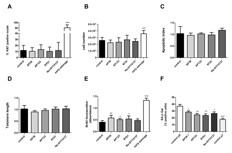
Figure 2.
Cell proliferation rate is not affected by H2S donor treatment. (A) Proliferation index was assessed for treated cells as assessed by Ki67 immunofluorescence (>400 nuclei counted per sample). (B) Cell counts following 24h treatment with Na-GYY4137 at 100 µg/ml, AP39, AP123, RT01 at 10 ng/ml. (C) Apoptotic index in senescent cells treated with inhibitors as determined by TUNEL assay. Data are derived from duplicate testing of 3 biological replicates. (D) Telomere length was assessed by qPCR in three biological and 3 technical replicates. (E) BrdU incorporation into cellular DNA. Relative BrdU incorporation was assessed in 3 biological replicates and was calculated by normalization of data to values corresponding to untreated (control) cells and are expressed as % BrdU incorporation. (F) Effect of 24h treatment with Na-GYY4137 at 100 µg/ml, AP39, AP123, RT01 at 10 ng/ml on accumulation of senescent cells over 2 passages in early passage cells (PD = 44). Mean+- SD of three independent experiment is shown. Statistical significance is indicated by *** p<0.001. Error bars represent the standard error of the mean.

