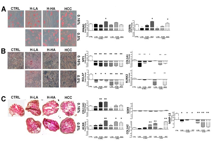
Figure 4.
Adipocyte (A), osteocyte (B), and chondrocyte (C) differentiation of MSCs treated with a different HA solution. On the left, there are representative images of Oil Red Oil (A), Alizarin Red S (B), and Safranin O (C) staining for every experimental condition. On the right, quantitative RT-PCR analysis of several differentiation markers. The mRNA levels were normalized to GAPDH mRNA expression, which was selected as an internal control. Histograms show expression levels in the different conditions. Data are expressed as arbitrary units with standard error (± SD, n = 3, *p < 0.05). For each gene in every experimental condition, the expression level in not-differentiated samples is set as the baseline (one value). Up- or down-regulation of genes in differentiated samples is shown as columns above or below the control baselines, respectively.

