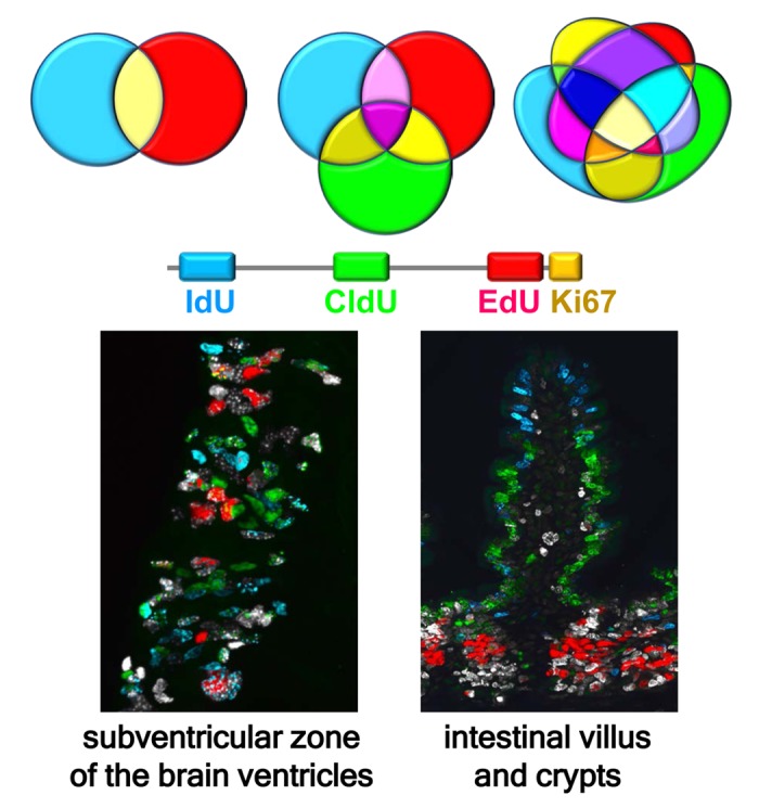
Figure 1.

Multiple labeling of dividing cells reveals defined subsets of stem and progenitor cells and their progeny. The number of differentially labeled subpopulations of cells expands exponentially as the number of individual tags and their possible combinations increases, even if only a minority of them correspond to biologically meaningful cell cohorts. Shown is an example of quadruple labeling of dividing cell cohorts in the subventricular zone (SVZ) of the lateral ventricles (left) and in the villi and crypts of the intestine (right). IdU, CldU, and EdU were sequentially injected in mice every 24 hr and the tissues analyzed 2 hr after the last injection, revealing three injected DNA tags and Ki67, an endogenous marker of dividing cells. Cells labeled with one, two, three, and four markers can be observed. Labeling of the brain shows currently or recently dividing neural stem and early progenitor cells and their progeny in the SVZ. Labeling of the intestine reveals rapid migration of newborn cells from the crypts to the top of the villi, with blue IdU-positive cells at the top of the villus, green CldU-positive cells in the middle segment of the villus, red EdU-positive cells strictly in the crypts, and white Ki67-positive cells in the crypts and the basal segment of the villus. Zones of non-overlapping colors reflect rapid ascent of enterocytes, born in the crypts, along the villi.
