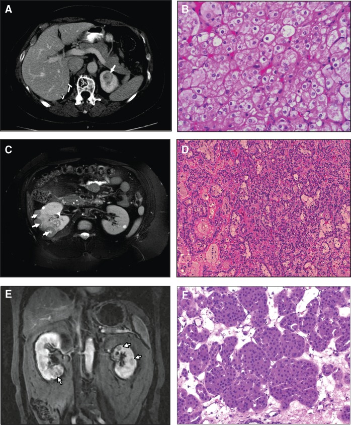
Figure 1.
Imaging and renal pathology of patients with FLCN H255Y and K508R mutations. Case 1: Patient from NCI Family with FLCN H255Y mutation. (A) MRI showing renal tumour in left kidney of patient with FLCN H255Y mutation. (B) Patient underwent a left partial nephrectomy in which 3 hybrid oncocytic tumours were removed. Magnification 200X. Case 2: Bilateral multifocal papillary type 1 RCC patient with FLCN K508R mutation. (C) MRI scan showing multiple renal tumours in right kidney of patient with FLCN K508R mutation. (D) This patient had a right partial nephrectomy in which 22 papillary type 1 renal tumours were removed. Magnification 150X. Case 3: Bilateral multifocal oncocytoma patient with FLCN K508R mutation. (E) MRI scan showing multiple renal tumours in right and left kidneys of patient with FLCN K508R mutation. (F) This patient had a left partial nephrectomy in which 5 renal oncocytomas were removed. Magnification 150X.

