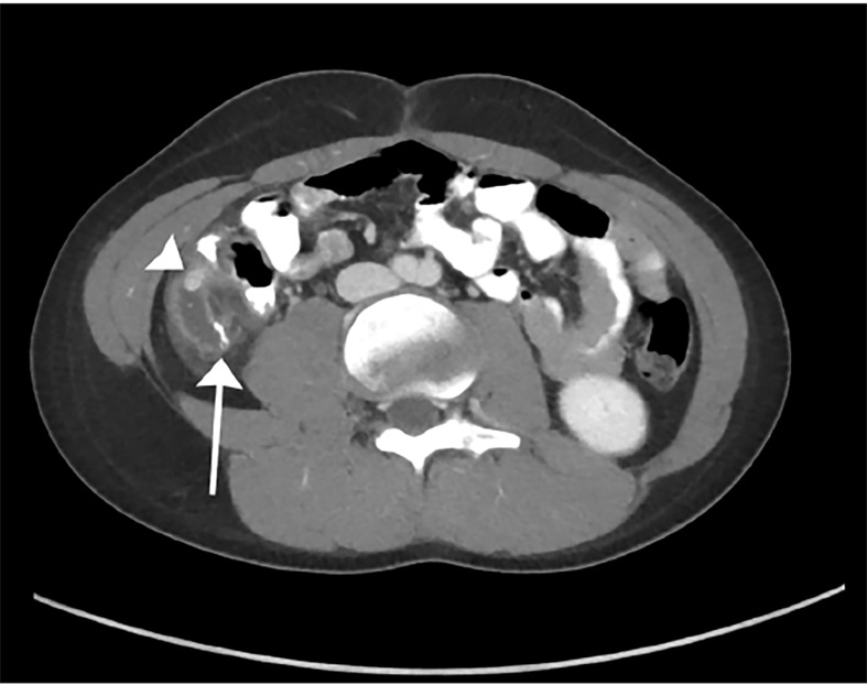
Image 1.
Contrast-enhanced computed tomography of the abdomen and pelvis, oblique axial plane, demonstrating surgical changes of appendectomy with staple lines at the blind end of the appendiceal stump (arrow). A high-density appendicolith (arrowhead) was obstructing the base of the appendiceal stump, which was surrounded by inflammatory changes.

