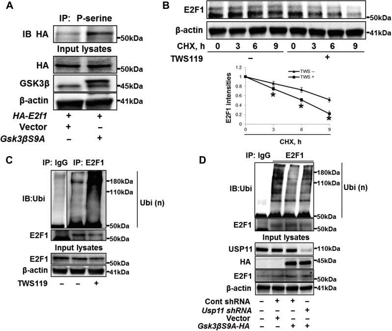
Figure 5.
GSK3β phosphorylates E2F1 and facilities E2F1 deubiquitination and stability. (A) A549 cells were transfected with vector or GSK3β mutant with S9A (Gsk3βS9A) for 24 h, and then transfected with HA-tagged E2F1 (HA-E2f1) plasmid for 24 h. Cell lysates were analyzed by IP. (B) A549 cells were treated with CHX (300 μg/ml) with or without TWS119 (10 μM) for 0, 3, 6, and 9 h. Cell lysates were immunoblotted with E2F1 and β-actin antibodies. E2F1 intensities were measured by ImageJ software. *P < 0.01, compared to TWS(−) treated cells. (C) A549 cells were treated with TWS119 (10 μM) for 2 h and analyzed by in vivo ubiquitination assay with a modified IP. (D) A549 cells were transfected with Cont shRNA, Usp11 shRNA, or Gsk3βS9A-HA for 48 h as indicated and analyzed by in vivo ubiquitination assay with a modified IP.

