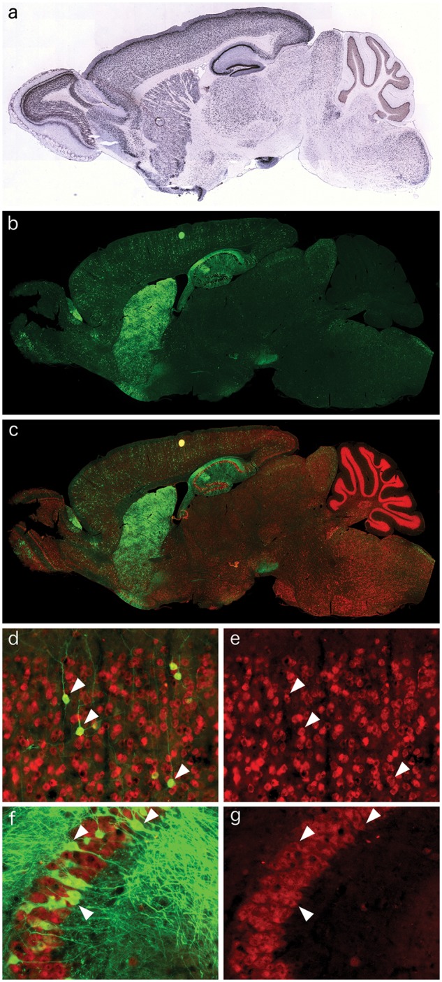
Figure 1.

Neuronal distribution of AAV9-CamKII-GFP in the Npc1-/- mouse brain after retro-orbital injection. (A) Excerpt from the Allen Brain Atlas, demonstrating the neuronal expression pattern of CamKII in the wildtype mouse brain. (B) Immunofluorescence of AAV9-CamKII-GFP (green) in the Npc1-/- brain after retro-orbital injection. (C–G) Co-localization of the AAV9-CamKII-GFP signal with NeuN immunofluorescence (red), indicating incorporation of AAV9-CamKII-GFP into neuronal populations, including cortical pyramidal neurons (D, green removed in E to show double labelling, arrowheads) and CA3 hippocampal neurons (F, green removed in G to show double labelling, arrowheads).
