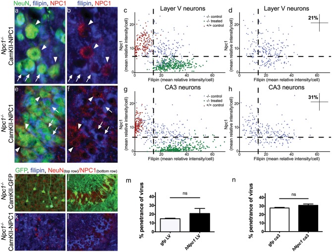
Figure 4.
Biochemical correction of the cholesterol storage phenotype in neurons transduced with AAV9-CamKII-NPC1 in the Npc1-/- mouse. Immunohistochemical imaging of NPC1 protein levels (red) in Layer V neocortex (A,B) and CA3 of hippocampus (E,F), co-stained with filipin (blue) and NeuN (green, in A and E only). Arrows indicate neurons without appreciable NPC1 protein and high filipin staining. Arrowheads indicate neurons successfully transduced by the AAV9-CamKII-NPC1 with subsequent intense NPC1 staining and reduced filipin labelling. (C,D) NPC1 intensity of all Layer V neurons measured, plotted against filipin intensity. (G,H) NPC1 intensity of all CA3 hippocampal neurons measured, plotted against filipin intensity. Upper left quadrants in (D,H) indicate the percentage of Npc1-/- neurons corrected to control levels with AAV9-CamKII-NPC1 treatment. Imaged density of AAV-CamKII-GFP (I,J) and AAV9-CamKII-NPC1 (K,L) incorporation in the Layer V cortex (I,K) and CA3 hippocampus (J,L), with quantification in (M,N). ns = non-significant.

