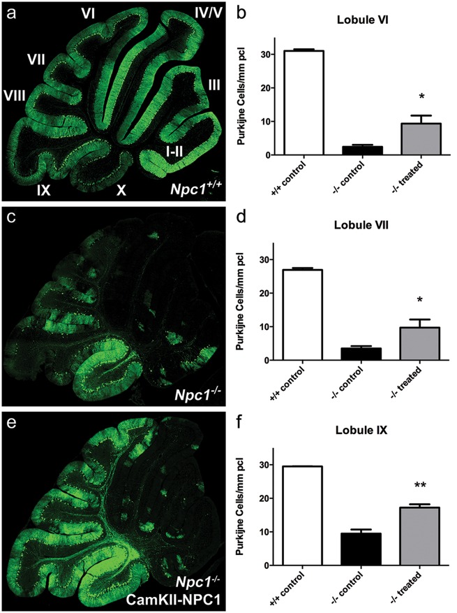
Figure 5.
Delayed Purkinje cell death upon AAV9-CamKII-NPC1 treatment in the Npc1-/- mouse. Immunofluorescent calbindin staining of Purkinje cells (green) in Npc1+/+ mice (A), Npc1-/- mice without treatment (C), and Npc1-/- mice treated with AAV9-CamKII-NPC1 (E) at 9 weeks of age [I-X in image (A) = cerebellar lobule number]. (B,D,F) Quantification of Purkinje cell number (positive cells determined by presence of staining in Purkinje cell body and dendritic arbor or axon) in posterior cerebellar lobules. All data in bar-graphs expressed as mean ± S.E.M., * P < 0.05, ** P < 0.01, one-way ANOVA with Tukey’s post-test.

