Figure 7.
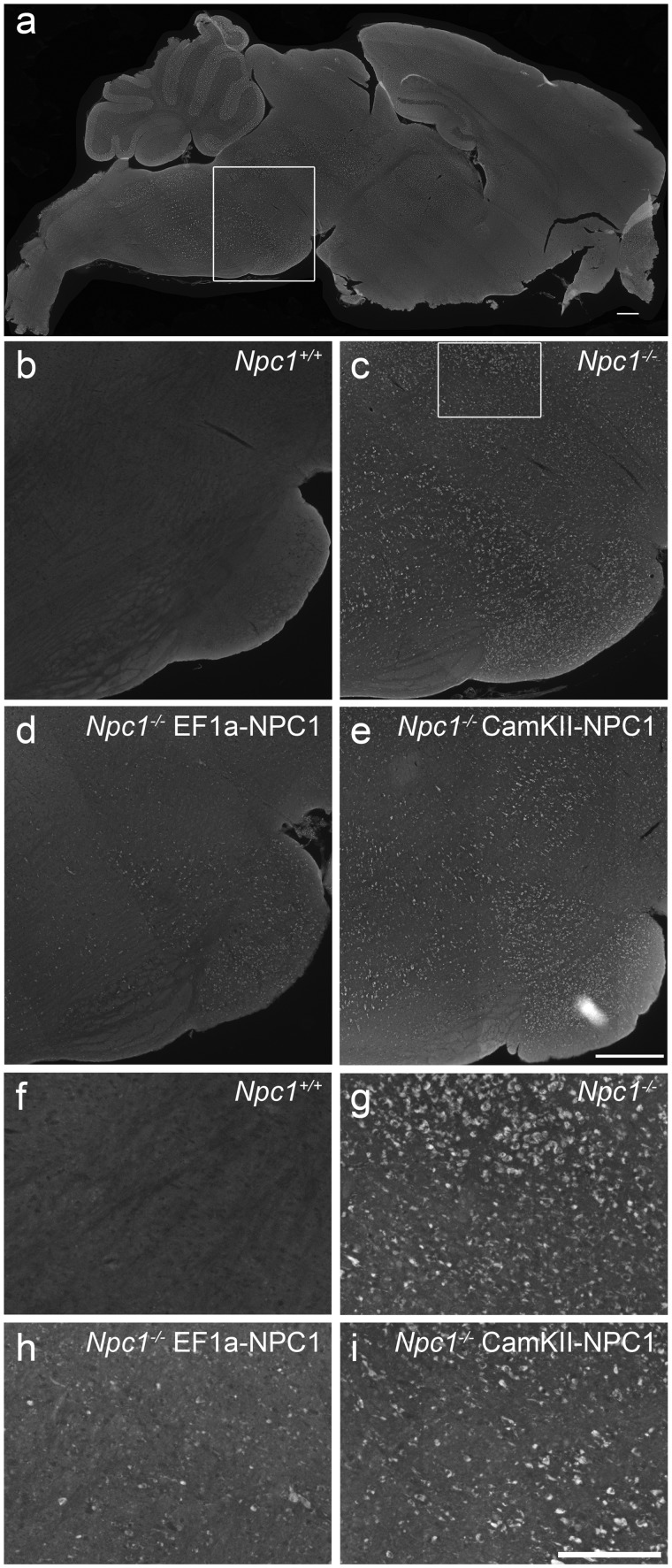
Effect of AAV9-EF1a-NPC1 vs AAV9-CamKII-NPC1 treatment in brain of Npc1-/- mice. (A–E) Immunohistochemical imaging of cholesterol (visualized with filipin labelling and noted as white intracellular punctae) in the pons, as outlined by box in (A). Npc1+/+ mice exhibit no cholesterol storage (B) while abundance of cholesterol laden vesicles are apparent in Npc1-/- mice (C). Npc1-/- mice treated with AAV9-EF1a-NPC1 exhibit reduced cholesterol accumulation (D) and Npc1-/- mice treated with AAV9-CamKII-NPC1 show intermediate correction of pathology (E). (F–I) Magnification of region outlined by box in (C) for figures (B–E) to facilitate easier visual comparison of cholesterol storage between treatment groups. Scale bars = 100 µm.

