Figure 8.
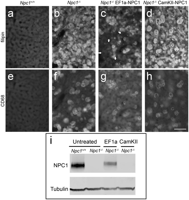
Effects of AAV9-EF1a-NPC1 vs AAV9-CamKII-NPC1 gene therapy on the liver of Npc1-/- mice. Immunohistochemical imaging of cholesterol (filipin, A–D) and Kupffer cells (CD68+, E–H). Npc1+/+ mice (A,E) exhibit no cholesterol accumulation or filipin staining (A) and Kupffer cells are minimal (E) whereas Npc1-/- mice without treatment (B,F) have extensive cholesterol accumulation (B) and increased abundance of Kupffer cells (F). AAV9-EF1a-NPC1 treated Npc1-/- mice (C,G) have hepatocytes free of cholesterol storage (C; e.g. white arrows) but demonstrate cholesterol storage in Kupffer cells (G), while AAV9-CamKII-NPC1 treated Npc1-/- mice show no correction of either cell type (D,H). Scale bar = 50 μm. (I) Western blot of liver homogenate from control and treated mice showing NPC1 protein (∼180 kDa) in the untreated Npc1+/+ and an AAV9-EF1a-NPC1 treated Npc1-/- mouse with Tubulin (50 kDa) serving as loading control.

