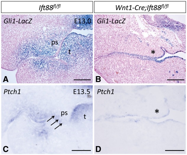
Figure 6.

Ciliary defects in Wnt1-Cre;Ift88fl/fl mice result in loss of Shh signaling in the palatal mesenchyme. (A, B) X-gal staining of E13.0 Ift88fl/fl;Gli1-LacZ (control, n = 3) and Wnt1-Cre;Ift88fl/fl;Gli1-LacZ mice (n = 3). Blue color indicates Gli1-LacZ positive cells. (C, D) In situ analysis of Ptch1 (blue staining) in E13.5 Ift88fl/fl(control, n = 6) and Wnt1-Cre;Ift88fl/flmice (n = 6). Arrows indicate the expression of Ptch1 in the mesenchyme. Asterisks indicate the downregulated expression of Gli1 and Ptch1 in the palatal mesenchyme. ps, palatal shelf; t, tongue. Scale bars, 200 µm.
