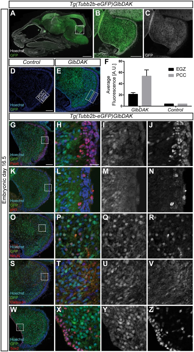
Figure 5.
Tubb2b is expressed at high levels in the embryonic cerebellum. (A–C) Overview scan of a sagittal section of an embryonic day (E) 16.5 head of the Tg(Tubb2b-eGFP)GlbDAK line. eGFP is expressed under the control of the endogenous Tubb2b locus on a bacterial artificial chromosome (BAC). (B) shows a magnification of the dashed rectangle encircling the cerebellar anlage in (A); in (C), the GFP channel is shown as a grey-scale image of (B). Hoechst staining for nuclear DNA is shown in white in A and B. (D,E) Confocal images of a control (D) and a transgenic animal (E). Hoechst staining for nuclear DNA is shown in blue in (D) and (E). PCC: Purkinje cell cluster area; EGZ: External Granular Zone. (F) Quantification of the average fluorescence intensity found in the EGZ and the PCC for control (n = 1) and transgenic mice (n = 3). For transgenic mice, mean ± SEM are shown. (G–Z) Fluorescent images of the developing cerebellum of the Tg(Tubb2b-eGFP)GlbDAK line at E16.5 stained for Pax6 (G–J), pH3 (K–N), NeuN (O–R), Calbindin (S–V) and Sox2 (W–Z). (H, L, P, T and X) show magnifications of the dashed rectangle in (G,K, O,S and W) respectively. (I, M, Q, U and Y), as well as (J, N, R, V and Z) show grey-scale images of the GFP and immunostaining channels, respectively. Scale bars show 1000 µm in A, 200 µm in B, 100 µm in (D) and (G) and 20 µm in (H).

