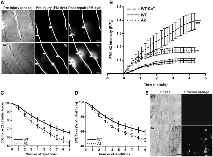
Figure 1.
Lack of AnxA2 causes poor sarcolemmal repair. (A) Freshly isolated intact Soleus (SOL) muscle isolated from WT or A2 mice were injured ex vivo in presence of FM1-43 dye. The images show time lapse images of fibers visualized for brightfield (pre injury, left panel), and for fluorescence emission of the FM dye pre and post injury (middle, right panels). Individual myofibers are marked by dotted white line, and arrows indicated the site of sarcolemmal injury. Scale bar = 20 μm. (B) Quantification of FM1-43 influx, following laser injury, into fiber isolated from WT in presence (n = 14 fibers) or absence of calcium (n = 4 fibers) and AnxA2 deficient (n = 22 fibers) mice. (C) Percentage of initial force as a result of repeated 10% ex vivo lengthening contractions of WT or A2 Extensor Digitorum Longus (EDL) muscle from 1 year old animal (n = 3–4 animals each). (D) Percentage of initial force as a result of repeated 10% ex vivo lengthening contractions of WT or A2 SOL muscle from 1-year-old animal (n = 3 animals each). (E) Cross-section of EDL muscle section stained with procion orange following 10 repeated lengthening contractions in WT and A2 from 1-year-old animal: brightfield (left panel) and procion orange (right panel). Scale bar = 50 μm. All images were acquired and scaled similarly and quantifications show means ± S.E.M. B: ** P ≤ 0.01 and ***P ≤ 0.001 compared to WT and ###P ≤ 0.001 compared to A2 by ANOVA; C: *P ≤ 0.05 compared to WT by unpaired t-test.

