Figure 3.
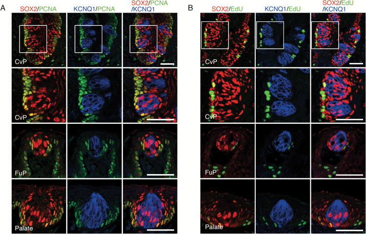
Identification of actively proliferating cells in the gustatory areas of oral epithelium. (A) Triple fluorescent labeling of SOX2 (red), PCNA (green), and KCNQ1 (blue) in the CvP (top and second-top), FuP (second-bottom), and the soft palate (bottom). Actively proliferating PCNA+ cells are observed in the basal epithelial cells outside taste buds, which are colabeled with SOX2. (B) Nucleoside analog (EdU) incorporation in actively proliferating cells in CvP (top and second-top), FuP (second-bottom), and soft palate (bottom): triple fluorescent labeling of EdU (green), SOX2 (red), and KCNQ1 (blue) at 4 h after EdU administration. Cells positive for both SOX2 and EdU are observed in the basal epithelium outside KCNQ1+ cells. Areas of magnified fluorescent images in row 2 (second-top) are indicated by white squares in row 1 (top). Scale bar: 50 µm.

