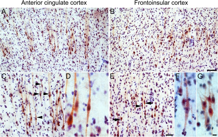
Figure 3.
VMAT2 immunohistochemistry in human ACC and FI Layer 5. Layer 5-predominant brown VMAT2 immunoreactivity is seen in the ACC (A) and FI (B) and prominently includes VENs and fork cells, as well as a subset of neighboring neurons with a pyramidal morphology (C, E). Higher magnification views of VMAT2-positive VENs in ACC (D) and FI (F) and fork cells in FI (G). Black arrowheads indicate examples of stained VENs, black arrows indicate stained fork cells, and white arrows indicate stained pyramidal-shaped neurons. Scale bars represent 100 μm (A, B), 50 μm (C, E), or 10 μm (D, F, G); apical at top.

