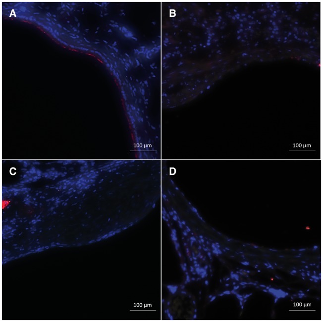
FIG. 3.
Immunofluorescent staining for common epithelial markers, including AT (red) and CCSP (green) of proximal bronchiolar epithelium (size bar: 100 µm). A, Naïve rat proximal bronchiolar epithelium with the majority of cells staining positive for AT but with a few, sporadic cells positive for CCSP. B, Two days after SM exposure, there was near complete loss of staining for both AT and CCSP. C, Fourteen days after SM injury, staining for ciliated and club cells remained absent with areas of complete denudation. D, Twenty-eight days after SM injury, staining of the proximal epithelium remained absent for AT and CCSP.

