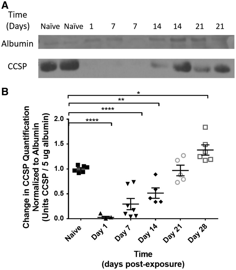
FIG. 5.
A, Western blot of BALF for CCSP quantification (bottom line). Albumin was used as the positive standard control (top line). B: Quantification of change in BALF CCSP normalized to albumin on postexposure day to amount of BALF CCSP normalized to naïve control. CCSP was significantly reduced on day 1 (0.03 ± 0.13 vs. 1.00 ± 0.03; P < .0001) and remained reduced on day 7 (0.29 ± 0.12; P < .0001) and day 14 (0.51 ± 0.13; P < .01) after SM exposure. By day 21 postexposure, CCSP returned to levels equivalent to that of naïve controls (0.97 ± 0.13; P > .05). By day 28 postexposure, BALF CCSP was increased in exposed animals compared with controls (1.38 ± 0.13 vs 1.00 ± 0.03; P < .05).

