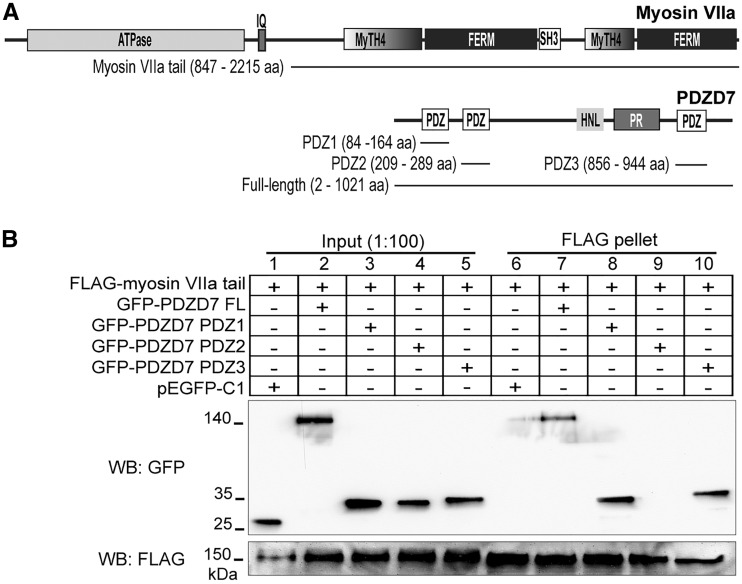
Figure 1.
Myosin VIIa interacts with PDZD7. (A) Domain organization of myosin VIIa and PDZD7. Lines below each diagram indicate the myosin VIIa and PDZD7 fragments used in this study. (B) FLAG-myosin VIIa tail pulled down GFP-PDZD7 full-length (FL, lane 7), PDZ1 (lane 8) and PDZ3 (lane 10) proteins, but not GFP-PDZD7 PDZ2 (lane 9) or GFP (pEGFP-C1, lane 6) protein. The lower anti-FLAG blot demonstrates the success of the FLAG pull-down assay. Lanes 1-5 are input samples showing the presence of the transfected proteins in cell lysates. + and -, presence and absence of proteins in the transfected cells, respectively.

