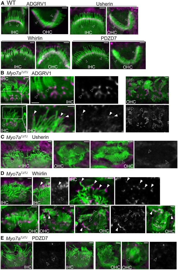
Figure 2.
USH2 protein distribution is abnormal in Myo7a7J/7J cochlear hair cells. (A) USH2 protein distribution in wild-type (WT) cochlear IHCs and OHCs at P4. ADGRV1, usherin, whirlin and PDZD7 (magenta) are localized to the ankle link region at the base (empty arrows) of stereocilia (phalloidin, green), while whirlin is also present at the stereociliary tip. The immunoreactive specificity of our USH2 antibodies in these stereociliary locations was previously verified by absence of their immunoreactivities in their corresponding mutant cochlear stereociliary bundles (15). Note that the magenta signals outside the stereociliary bundle are non-specific. For an unknown reason, our usherin antibody usually gave this non-specific signal. (B) In P4 Myo7a7J/7J IHCs and OHCs, a large amount of ADGRV1 forms aggregates at the medial side of the stereociliary bundle on the apical surface of hair cells, while a trace amount of ADGRV1 remains at the ankle links (empty arrows) and is mislocalized at the stereociliary tips (filled arrows). The signal intensity at the stereociliary tip was ∼10% of those at the stereociliary base and at the cell apex and was thus enhanced in order to be seen. Boxed regions are amplified and shown on the right of the original images. n ≥ 2 pups from 1 litter. (C) Usherin immunofluorescence is undetectable in P4 Myo7a7J/7J IHCs and OHCs. Empty arrows: the base of stereocilia. n ≥ 3 pups from 3 litters. (D) Immunofluorescent signals of whirlin are detectable at the tip (filled arrows) but not at some of the ankle link region (empty arrows) in P4 Myo7a7J/7J cochlear stereociliary bundles. n ≥ 3 pups from 3 litters. (E) Immunoreactivity of PDZD7 is barely detectable at the stereociliary base (empty arrows) of P4 Myo7a7J/7J IHCs and OHCs. n ≥ 2 pups from 1 litter. Some single-channel images of USH2 proteins are shown in grayscale on the right of or below the merged images, where the position of arrows is matched in the single-channel and merged images. Scale bars, 1 µm.

