Figure 3.
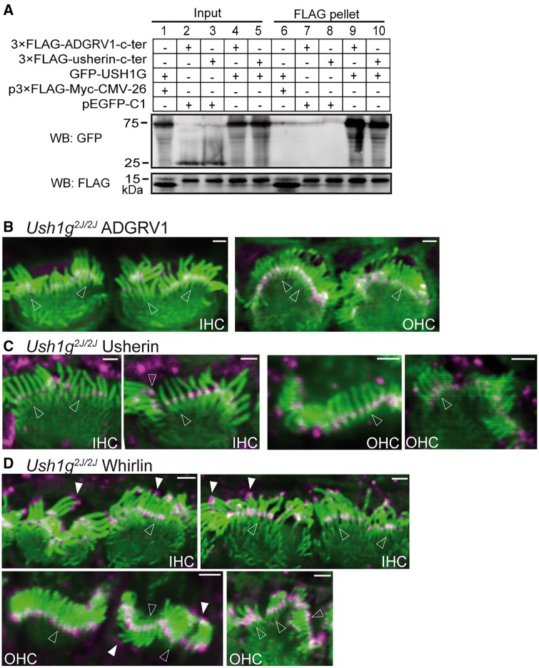
Interactions of USH1G with usherin and ADGRV1 and distribution of USH2 proteins in Ush1g2J/2J cochlear hair cells. (A) GFP-USH1G (lane 9 and lane 10), but not GFP (pEGFP-C1, lane 7 and lane 8), was pulled down by FLAG-usherin and FLAG-ADGRV1 cytoplasmic fragments (c-ter) using anti-FLAG M2 affinity agarose. The FLAG peptide expressed by p3xFLAG-Myc-CMV-26 vector was used as a negative control (lane 6). The bottom anti-FLAG blot demonstrates the success of the FLAG pull-down assay. Lanes 1-5 are input samples showing the expression of GFP-USH1G, GFP, FLAG-usherin c-ter, FLAG-ADGRV1 c-ter and FLAG peptide in the transfected HEK293 cells. + and -, presence and absence of protein fragments in the transfected cells, respectively. (B-D) Although the stereociliary bundle (phalloidin, green) is significantly distorted in Ush1g2J/2Jcochlear hair cells, localizations of ADGRV1 (magenta, B), usherin (magenta, C) and whirlin (magenta, D) at the ankle link region (empty arrows) are normal. Additionally, whirlin localization at the stereociliary tips (filled arrows, D) is also normal in P4 Ush1g2J/2Jcochlear hair cells. n ≥ 2 pups from 2 litters for ADGRV1 immunostaining; n ≥ 4 pups from 4 litters for usherin immunostaining; and n ≥ 2 pups from 2 litters for whirlin immunostaining. The magenta signals outside the stereociliary bundle are non-specific. Scale bars, 1 µm.

