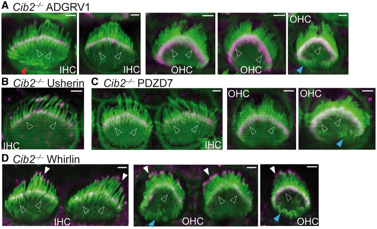
Figure 5.
Normal distribution of USH2 proteins in Cib2-/- cochlear hair cells. Immunofluorescent staining demonstrated that ADGRV1 (magenta, A), usherin (magenta, B), PDZD7 (magenta, C) and whirlin (magenta, D) are localized at the base (empty arrows) of stereocilia (phalloidin, green), while whirlin is also localized at the stereociliary tips (white filled arrows) in P4 Cib2-/- cochlear hair cells. Blue and red arrows point to the ectopic stereocilia found in Cib2-/- OHCs and IHCs, respectively. Experiments were performed in 3 litters of Cib2-/- pups for ADGRV1 immunostaining, 1 litter for usherin immunostaining, 1 litter for PDZD7 immunostaining and 2 litters for whirlin immunostaining. Scale bars, 1 µm.

