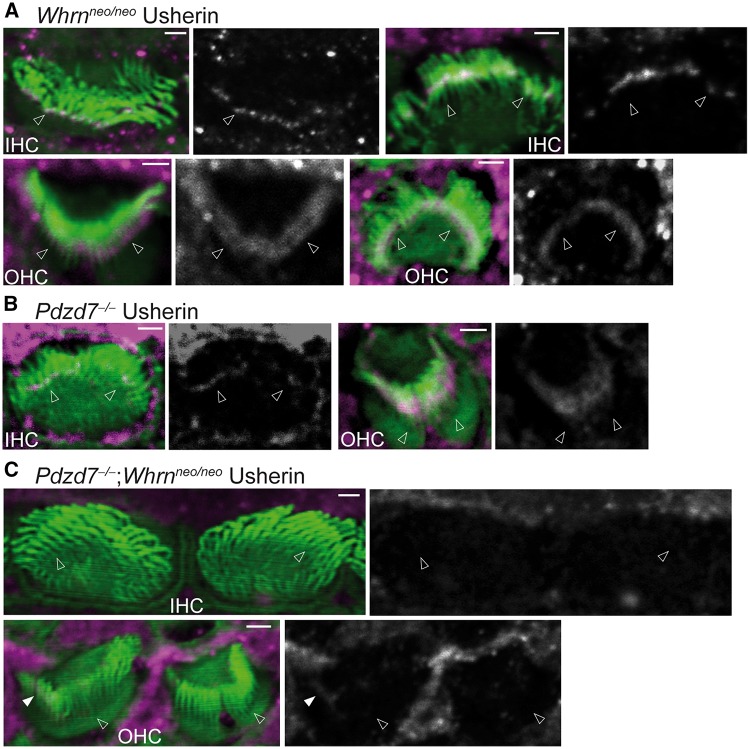
Figure 7.
Usherin is undetectable at the ankle link region in the Pdzd7-/-;Whrnneo/neo cochlear stereociliary bundle. (A) Usherin (magenta) is normally localized at the base of stereocilia (phalloidin, green) in P4 Whrnneo/neo IHCs and OHCs. n ≥ 3 pups from 3 litters. (B) Usherin is reduced at the stereociliary base of Pdzd7-/- IHCs and moves toward stereociliary tips in Pdzd7-/- OHCs at P4. Usherin signal was enhanced in the IHC to show the reduced signal at the stereociliary base. n ≥ 3 pups from 3 litters. (C) Usherin immunoreactivity is absent in the IHC stereociliary bundle and is barely detectable in the OHC stereociliary bundle in P4 Pdzd7-/-;Whrnneo/neo cochleas. The filled arrow points to an extremely weak and diffuse usherin signal in the middle of stereocilia. n ≥ 2 pups from 2 litters. The magenta signals outside the stereociliary bundle in this figure are non-specific and were usually seen for usherin staining. Single-channel images of usherin signals are shown in grayscale on the right of the merged images with the position of arrows the same as that in the merged images. Empty arrows point to the base of stereocilia. Scale bars: 1 μm.

