FIG. 3.
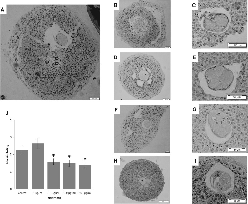
Effect of the phthalate mixture on antral follicle atresia. Antral follicles were isolated from mouse ovaries and treated with vehicle (DMSO) or phthalate mixture (1–500 µg/ml) for 96 h. Following culture, antral follicles were processed for histological evaluation of atresia. A representative image of a DMSO treated follicle is found in panel A, representative apoptotic bodies are circled. Representative images of mixture at 1, 10, and 100 μg/ml with intact oocytes are shown in B, D, and F, respectively. Representative images of mixture at 1, 10, and 100 μg/ml with fragmented oocytes are shown in C, E, and G, respectively, Panel H and I are images of oocytes in the 500 μg/ml treatment group. All scale bars indicate 50 μm. Atresia scores are shown in panel J. A score of 1 indicates the presence of apoptotic bodies encompassing 0–3% of the total area of follicle, a score of 2 indicates the presence of apoptotic bodies encompassing 4–10% of total area of follicle, a score of 3 indicates the presence of apoptotic bodies encompassing 11–30% of total area of follicle, and a score of 4 indicates >30% of apoptotic bodies of the total area of follicle. Graph represents means ± SEM from 3 separate experiments, with 12 follicles/treatment group in each experiment. Asterisks (*) represent significant difference from the control (P < .05).

