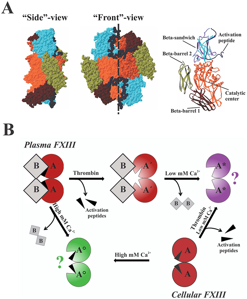
Fig. 1. Structure and activation of the A-subunit of FXIII.

(A) Zymogen FXIIIA2 (PDB 1F13) is formed by two identical subunits oriented in a head-to-tail fashion (shown as space-filling models, dashed line denotes homodimer interface), with the activation peptide of each subunit blocking access to the active site of the neighboring subunit. Structural domains of the left subunit are highlighted in a ribbon representation. (B) Both plasma and cellular FXIII can be activated by thrombin in the presence of low mM Ca2+ or non-proteolytically by high (>50 mM) concentration of Ca2+. Aʹ - FXIIIA after cleavage of the activation peptide; A* - FXIIIA activated by thrombin in the presence of Ca2+; A° - non-proteolytically activated FXIIIA; question marks denote unknown oligomerization state. A more detailed description can be found in the text.
