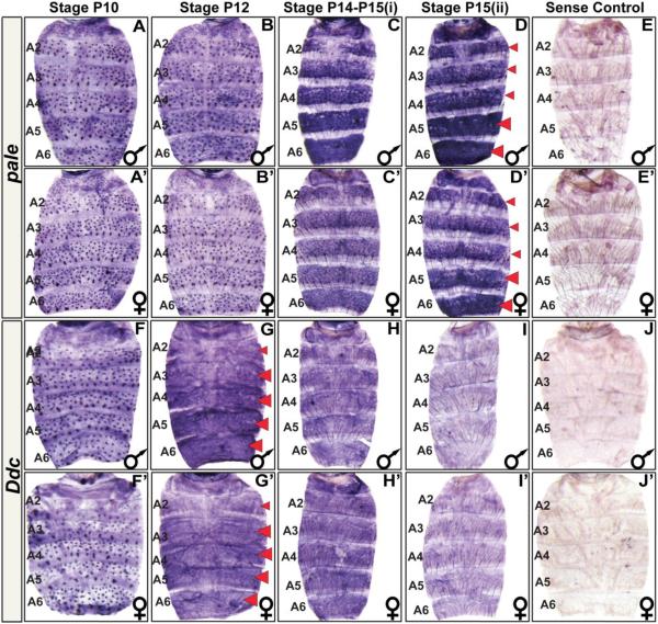
Fig. 2. The spatial and temporal expression pattern of D. melanogaster pale and Ddc.
(A–J) Samples for D. melanogaster males and (A’–J’) females. (A and A’) pale is expressed in the mechansosensory bristle cells as early as the P10 stage of pupal development. (B and B’) Mechanosensory cell expression continues through the P12 stage. (C and C’) By the P14-P15(i) stage, pale expression has dramatically increased in the abdominal epidermis. Most notably in the A5 and A6 segments and in the epidermal cells underlying where tergites will form. (D and D’) The epidermis pattern of pale expression can still be detected in newly eclosed flies. (E and E’) A sense probe control shows the background pattern of signal observed in an eclosed fly. (F and F’) Ddc is expressed in the mechansosensory bristle cells as early as the P10 stage of pupal development. (G and G’) By the P12 stage, Ddc expression has dramatically increased in the abdominal epidermis, most notably cells in the A3–A6 segment regions underlying where tergites will form. (H and H’) By the P14-P15(i) stage, Ddc expression persists but at an apparently reduced level. (I and I’) Epidermis expression has become difficult to detect in newly eclosed flies. (J and J’) A Ddc sense probe control shows background signal observed in eclosed fly. Red arrowheads indicate stages and segments with conspicuous expression detected in the epidermis. These stages were prioritized for investigations of expression in related fruit fly species.

