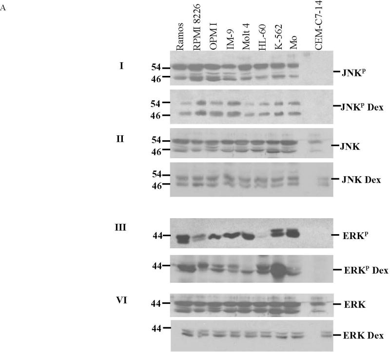
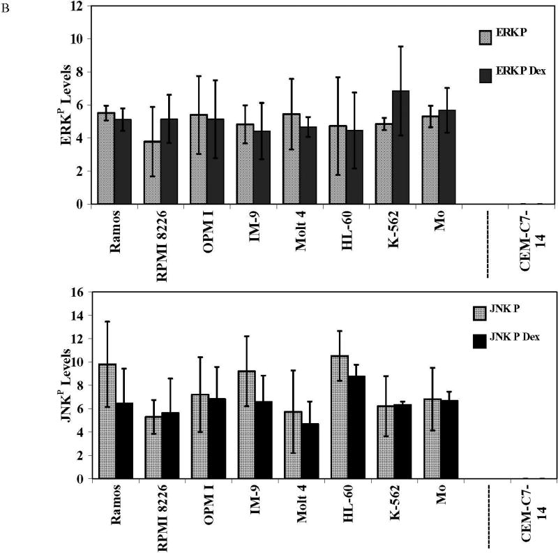
Fig. 1. All the Dex-resistant cell lines have high levels of JNKP and ERKP relative to Dex-sensitive CEM-C7-14 cells.
Cells were plated at 3×10⁵ viable cells/ml. After exposure to 1 µM Dex or vehicle for 24 h, cell lysates were prepared and analyzed by immunoblot for total and phosphorylated ERK and JNK. Each filter was subsequently blotted for β-actin (not shown). A. Characteristic blots from two experiments, Experiment 1, filters I and II, for phosphorylated (JNKP) and total JNK, each ± Dex. Experiment 2, filters III and IV, for ERKP and total ERK ± Dex. B. Average JNKP and ERKP levels, ± Dex, each cell line. Means from 3 independent experiments. Images were analyzed densitometrically and normalized to β-actin. Ordinate: Ratio of densitometry units, MAPK/actin. Error bars =1 standard deviation of average n=3 independent experiments; p value based on two-tailed students t-test using Excel.


