Figure 4.

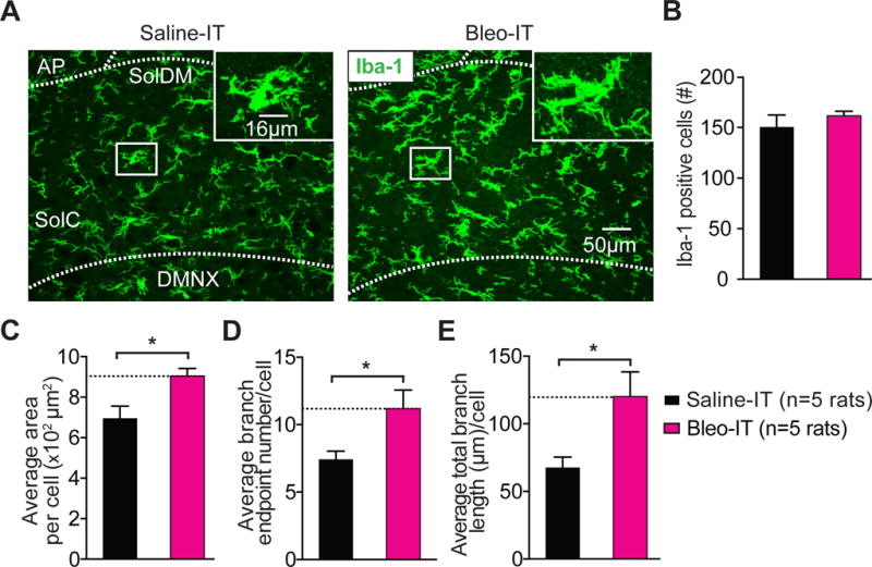
Lung injury in pre-transition period rats-pups promotes hyper-ramification of Iba-1+ microglia in the nTS and IL-1β/pro-IL-1β production throughout the dorsomedial brainstem.
A Representative images of coronal brainstem sections from saline-treated (black) and Bleo-treated (magenta) groups (7-days after injury) showing immunohistochemical staining for Iba-1+ cells (microglia) in the commissural (SolC), and dorsomedial (SolDM) subnuclei of the nTS. Inset: magnified images showing microglia localized to a medial region of the nTS.
B The number of microglia in the nTS was not significantly different in Bleo-treated (n = 5) or saline-treated (n = 5) rats (P = 0.411, Mean ± SEM, Two-tail t-Test).
C The area of individual microglia from Bleo-treated rats was greater than that of saline-treated rats (*P = 0.018, Two-tail t-Test).
D The number of branch endpoints per microglia increased in Bleo-treated compared to saline-treated rats (*P = 0.038, Two-tail t-Test).
E The total branch length per microglia increased in Bleo-treated compared to saline-treated rats (*P = 0.03, Two-tail t-Test).
