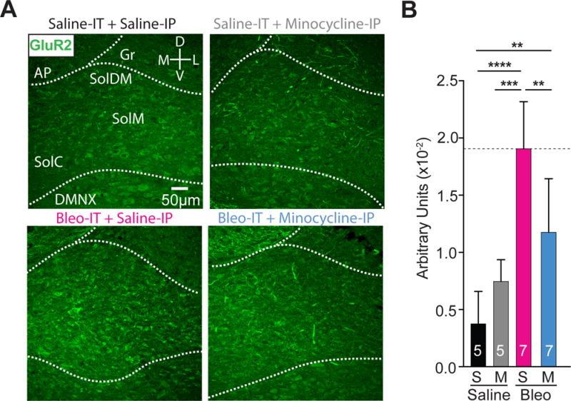
Figure 9.

Minocycline treatment in pre-transition period lung-injured rat-pups reduces GluA2+ immunostaining in the nTS
A Representative images showing coronal brainstem sections from saline-IT + saline-IP (S+S, n = 5) saline-IT + minocycline-IP (S+M, n = 5), Bleo-IT + saline-IP (B+S, n=7) and Bleo-IT + minocycline-IP (B+M, n = 7) treated groups immunostained for GluA2 in commissural (SolC), medial (SolM) and dorsomedial (SolDM) subnuclei of the nTS.
B GluA2+ staining in the nTS was significantly reduced in B+M (blue) treated rats compared to B+S (magenta) treated rats (**P = 0.008), but significantly greater than S+S (black) treated rats (**P = 0.0079), and was not significantly different from S+M (grey) treated rats (P = 0.234, One-way ANOVA with Tukey multiple comparisons test). There were no significant differences between S+S and S+M treated groups (P = 0.419).
