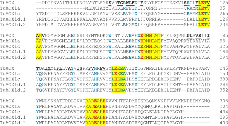
Fig 12. Alignment of TuAOX (T. urartu) proteins with TbAOX (T. brucei).
Yellow highlights indicate conserved motifs. Red font indicates residues proposed to coordinate the diiron center of the active site. Blue font indicates residues experimentally tested for loss of activity by previous researchers. Underlined residues are involved in the TbAOX hydrophobic cavity.

