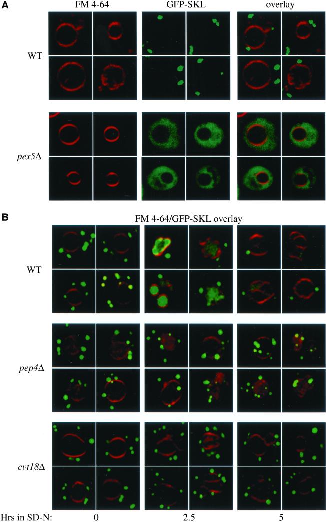
Figure 6.
Pexophagy is blocked at a prevacuolar stage in the cvt18Δ mutant. (A) GFP-SKL chimera is localized to peroxisomes. Cells from Wild-type (WT, WCG4a) and pex5Δ (MGY103) strains expressing GFP-SKL from a CEN plasmid were grown to mid log phase in SMD as described in MATERIALS AND METHODS, and observed on a scanning confocal microscope. The vital dye FM 4-64 was added to cultures to allow visualization of the vacuolar membrane. (B) Peroxisomes accumulate outside of the vacuole during pexophagy in the cvt18Δ mutant. Cells from WT (WCG4a), pep4Δ (YMTA), and cvt18Δ (JGY9) strains were grown to mid log phase in SMD and transferred to YTO to induce peroxisome proliferation, and subsequently transferred to SD-N to induce degradation of the excess peroxisomes as described in MATERIALS AND METHODS. Cells were examined by scanning confocal microscopy after incubation in SD-N at the indicated times.

