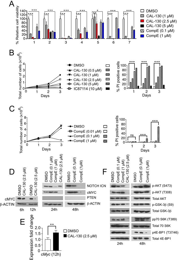
Figure 3.

PI3Kγ/δ inhibition promotes tumor cell death irrespective of NOTCH1 activation status. A, Analyses of primary CD2-Lmo2 mouse T-ALL tumor cell viability in the presence of increasing concentrations of CAL-130 or CompE for 72 h. B-C, Proliferation and survival analysis of a representative Lmo2-driven T-ALL tumor cell line (03007) cultured in the presence of increasing concentrations of CAL-130 (B) or CompE (C) for 72 h. Data represent mean ± SEM (n = 3 in triplicate). Statistical significance was determined by Student’s t test for drug treated cells relative to control. *, P < 0.01; **, P < 0.001; ***, P < 0.0001; ns (not-significant). D, Immunoblots of PTEN and cMYC expression as well as NOTCH1 activation state in a representative CD2-Lmo2 T-ALL cell line (03007) cultured in the presence of CAL-130 (6, 12, and 24 hours) or CompE (24 and 48 hours). E, Expression levels of cMyc in the identical tumor cell line after 12 h of treatment with CAL-130 (2.5 μM) as determined by qRT-PCR (n = 3). F, Immunoblots depicting AKT and the activation state of its downstream effectors in the same cell line at the indicated duration of treatments.
