Figure 5.
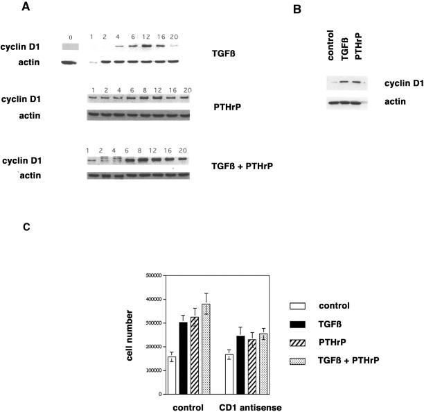
Induction of cyclin D1 expression by TGFβ and PTHrP. (A) RCS cells were serum-starved for 3 d, stimulated with control medium, TGFβ (1 ng/ml), PTHrP (10−8 M), or both for the indicated intervals, and harvested for the analyses of cyclin D1 protein expression by Western blot. Equal gel loading (5 × 10⁵ cells/lane) was documented by probing for actin protein expression. (B) Primary rat chondrocytes were serum-starved for 1 d, stimulated with control medium, TGFβ (1 ng/ml), or PTHrP (10−8 M) for 8 h, and harvested for the analyses of cyclin D1 protein expression by Western blot. Equal gel loading (5 × 10⁵ cells/lane) was documented by probing for actin protein expression. (C) Primary rat chondrocytes were plated in 24-well plates (105 cells/well). The cells were incubated with control or cyclin D1 antisense oligonucleotides and stimulated with control medium, TGFβ (1 ng/ml), PTHrP (10−8 M), or both for 3 d, and then counted with the use of a hemacytometer.

