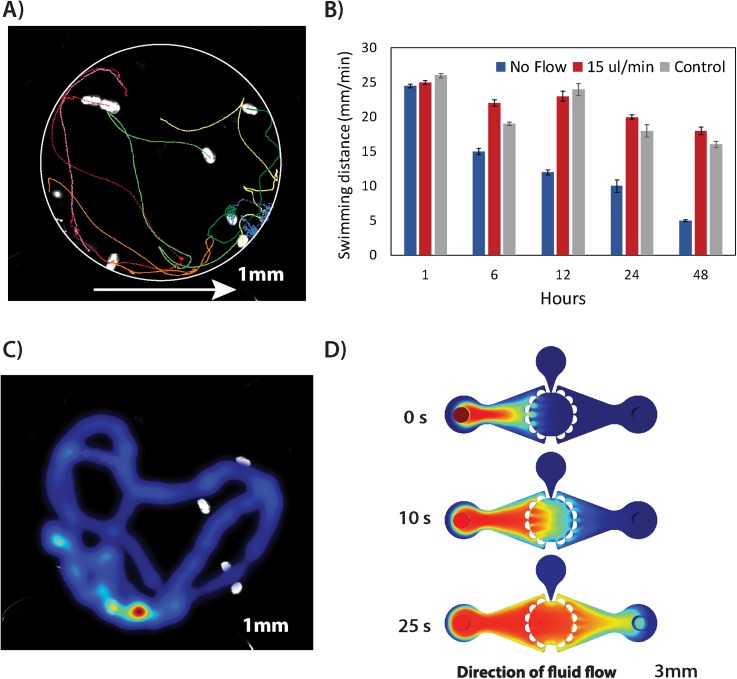
FIG. 2.
Validation of microfluidic environment for minimally invasive perfusion culture of caged rotifers. (a) Reconstruction of animal 2D trajectories in a caging test chamber when perfused at a volumetric flow rate of 15 μl/min. The plot denotes an average distance travelled [mm] of ten specimens caged in a microfluidic device. The white circle is a superimposed outline of the trapping chamber; arrow indicates a direction of the fluid flow. (b) Comparative analysis of rotifers' swimming behaviour (mean distance travelled ± SE) kept under different fluidic conditions. (c) Occupancy map depicting cumulative regions of animal presence in x-y space during 60 s of video when perfused at a volumetric flow rate of 15 μl/min. (d) Computational fluid dynamics (CFD) simulations of a mass transfer inside the chip-based device actuated at a flow rate of 15 μl/min.

