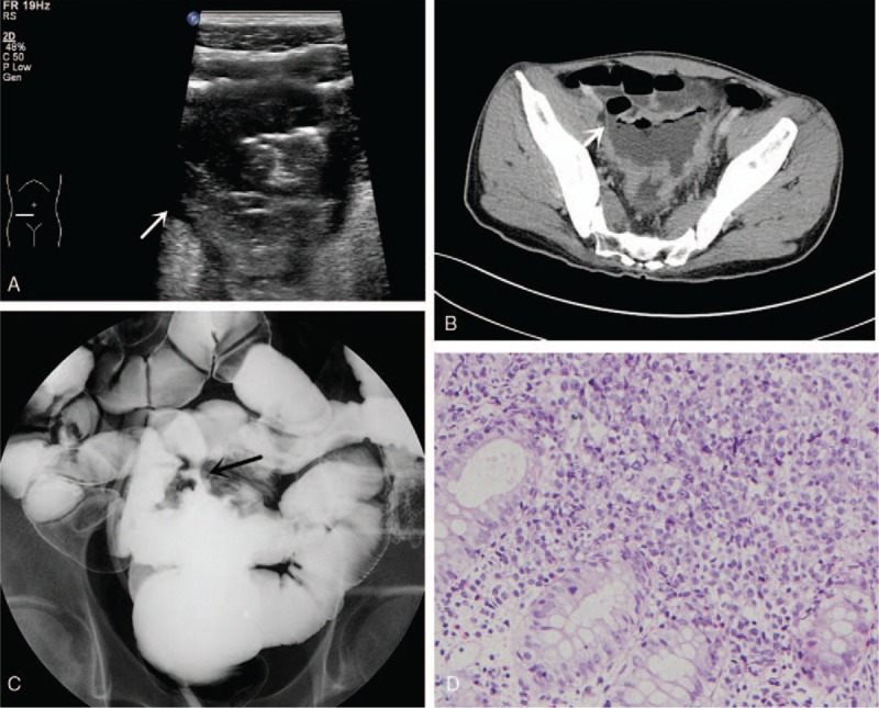
Figure 2.

(A) US shows the sigmoid colon adhered to the pelvic small intestine, a fistulous communication (white arrow) in which intestinal content that is moving can be seen between them. (B) CTE demonstrating that the wall of the partial sigmoid colon was abnormally thickened and kept a close relationship with the small intestine, suggesting an intestinal fistula (white arrow). (C) Barium enema showed a tract (black arrow) between the small intestine and the sigmoid colon. (D) Histopathologic result showed non-Hodgkin's large diffuse B-cell lymphoma.
