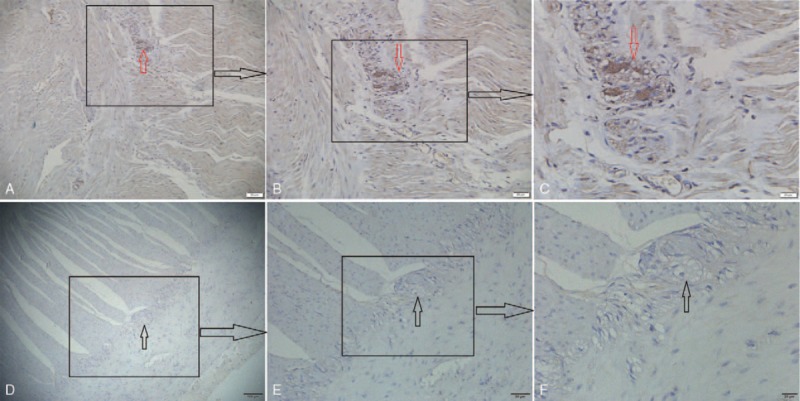
Figure 2.

The α-synuclein immunoreactivity in sections of intestinal submucosal intramuscular nerve plexuses. Expression of α-synuclein in the intramuscular nerve plexus of the intestinal submucosa, PD group: ABC (positive: red arrow), control group: DEF (negative: black arrow) (A × 100, B × 200, C × 400, D × 40). ABC: Distinct α-synuclein expression increased markedly in the submucosal intramuscular nerve plexuses, as the immunoreactive materials of α-synuclein were granularly distributed in the intramuscular nerve plexuses, mainly in the cytoplasm of the intramuscular ganglions, though a few granular positive structures were also scattered in the nerve fibers. DEF: No distinct α-synuclein expression increased markedly in the submucosal intramuscular nerve plexuses.
