
Figure 4.
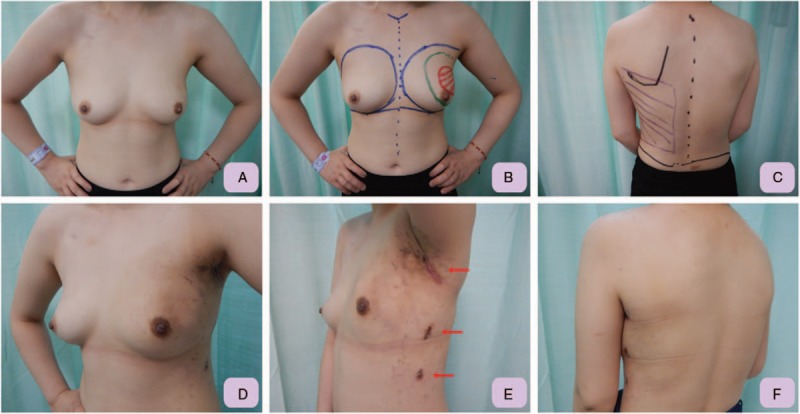
Pre- and post-operative pictures for patient who received RAQ and IPBR with RLDFH. A, Pre-operative front view of the 28 years old female with large left breast cancer post neoadjuvant chemotherapy indicated for left upper outer quadrantectomy. B, The location and presumed resection margin of left breast cancer was marked pre-operatively under the guidance of sonography. C, The borders of the LD muscle are marked: the anterior border was marked during LD muscle contraction, the superior border is marked over the tip of the scapula, and the posterior border is defined about 3 to 4 cm lateral to the spine. D, Left lateral-front view, which was taken 3 weeks post operation, showed that after IPBR with RLDFH, the left breast was in good shape and in symmetry with the right breast. E, Post-operative lateral view showed that the wound was small and well hidden in the inconspicuous axilla region. Two other 1 cm trocar wounds, which were used as drain exit sites, were also small and inconspicuous. F, Post-operative posterior view showed no incision scar was left over the back. IPBR = immediate partial breast reconstruction, LD = latissimus dorsi, RAQ = robotic assisted quadrantectomy, RLDFH = robotic latissimus dorsi flap harvest.