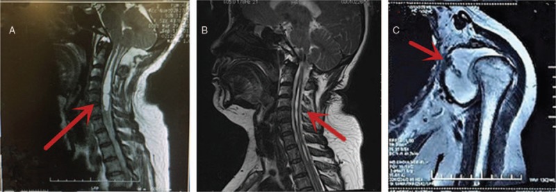
Figure 1.

(A) A sagittal T2-weighted magnetic resonance imaging scan of the cervical spine demonstrates a syrinx extending from the C2 vertebral body down to the T2 vertebral body; (B) 6-month follow-up after foramen magnum decompression shows the decrease in size of syrinx. (C) Magnetic resonance imaging of the left shoulder joint showing the shoulder joint disclosed and the glenoid losing its normal shape. A large amount of effusion can be seen in the joint cavity (arrow).
